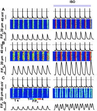
Figure 2.
Disturbances in rhythmic [Ca2+]i transients and membrane potential induced by ISO in myocytes expressing CASQ2D307H.
Recordings of membrane potential (upper traces), along with line scan images (middle) and time-dependent profiles of [Ca2+]i (lower traces) in myocytes infected with Ad-Control (A), Ad-CASQ2WT (B), and Ad-CASQ2D307H (C) vectors before and after exposure of the myocytes to 1 μmol/L ISO. Myocytes were stimulated at 2 Hz in the current-clamp mode. Reprinted from Viatchenko-Karpinski et al.12 with permission.