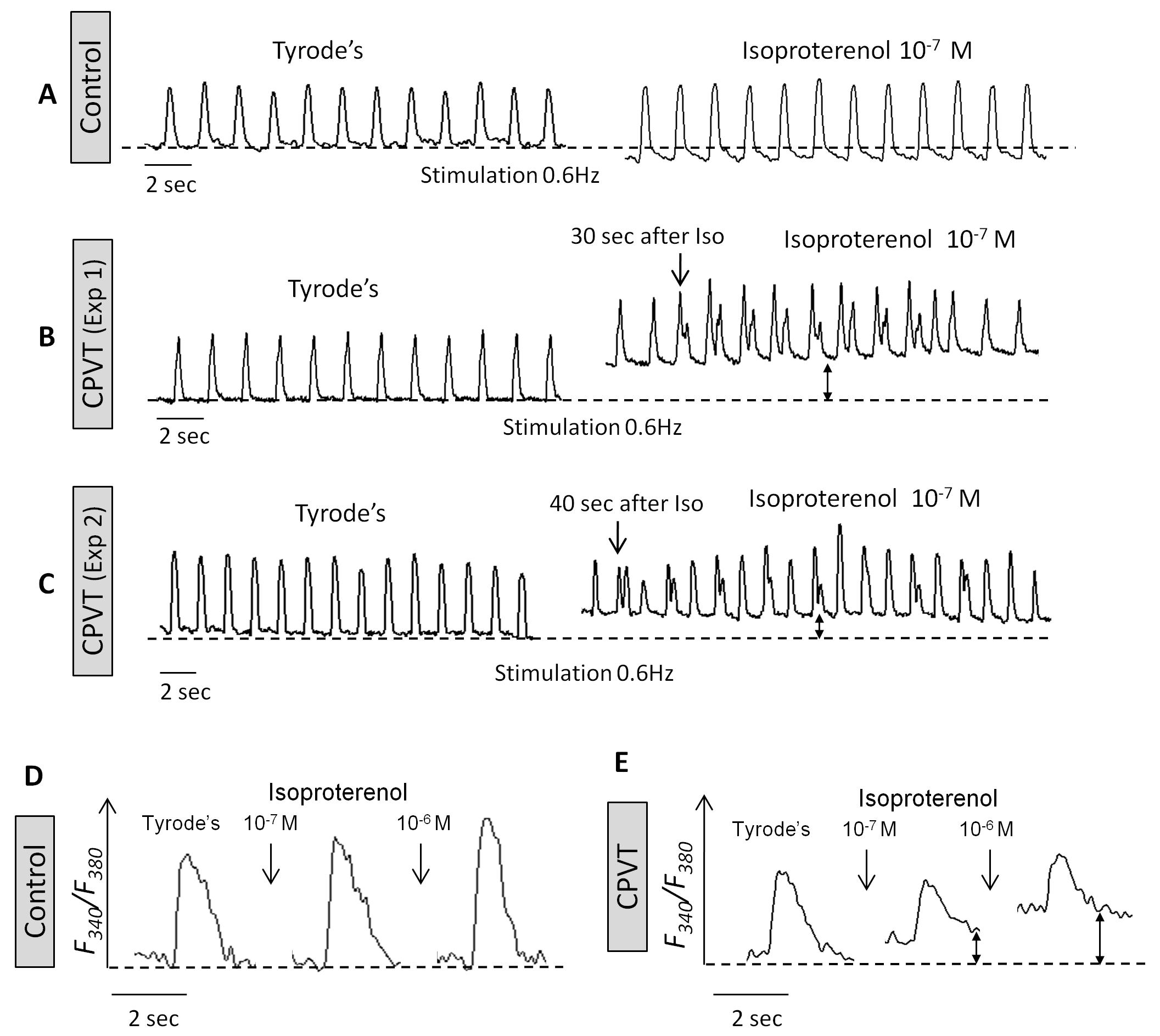
Figure 4.
The effects of isoproterenol on the [Ca2+]i transients and contractions in control and CPVT iPSC cardiomyocytes.
(A) Representative contraction tracings of control iPSC cardiomyocytes (43-day-old embryoid body (EB)) stimulated at 0.6 Hz, in the absence (Tyrode’s) and presence of isoproterenol. (B,C) Representative contraction tracings of CPVT iPSC cardiomyocytes (33- and 38-day-old EBs, respectively) stimulated at 0.6 Hz, in the absence (Tyrode’s) and the presence of isoproterenol. Note that after-contractions developed only in the CPVT cardiomyocytes in the presence of isoproterenol. (D,E) Representative [Ca2+]i transients of control iPSC cardiomyocytes (40-day-old EB) and CPVT iPSC cardiomyocytes (34-day-old EB), respectively, before and 5 minutes after isoproterenol perfusion. Reprinted from Novak et al.37 with permission.