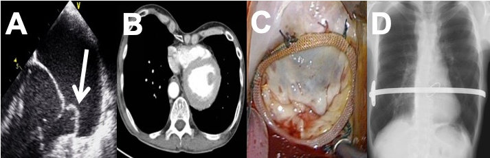
Figure 1. Perioperative and Procedural Images of Staged Endoscopic Mitral Valve Repair and Pectus Excavatum Correction Surgery
A: Trans-thoracic echocardiography indicating a prolapsing P2 mitral valve segment (white arrow). B: Computerized tomography indicating cardiac displacement to the left hemithorax. The Haller Index* and the Correction Index† were calculated to be 3.1 and 33.9%, respectively. C: Endoscopic mitral valve repair. D: Post-operative chest X-ray indicating satisfactory Nuss bar positioning.
*Haller Index: the ratio of the transverse thoracic (25.4 cm) and minimum sternovertebral diameter (7.6 cm).
†Correction Index: the indentation depth (3.9 cm) as a percentage of the maximum sternovertebral diameter (11.5 cm).